Microscopy Laboratory
Microscopy Laboratory
Electron microscopy has become ubiquitous in material-related analysis in academia, industry, medicine, and forensic. There are two units of scanning electron microscope (SEM) operating at the chemistry department. Tungsten filament SEM from Emcrafts and Schottky field emission SEM from ThermoFisher. Both SEMs are equipped with energy dispersive X-ray analysis (EDX) detector for elemental analysis.
Our two SEMs are able to provide topological and elemental analysis. These topological and elemental information originate mainly from three signals: secondary electrons (SE), back-scattered electrons (BSE) and characteristic X-rays. The proportion of the three signals detected by the electronics in the SEMs depends on the interaction between the primary beam (the stream of electrons coming from the electron gun) and the sample being investigated. This means the type of information generated depends on how energetics the electron beam is and on the texture and elemental composition of the sample.
In general SE signal is sensitive to the sample’s topology, giving information from the sample’s surface. BSE signal is sensitive to the sample’s composition based on Z-contrast. And finally elemental analysis from the characteristic X-ray signals. As a consequence of where the signals coming from (see figure for e-beam and sample interaction volume) the spatial resolution increases from X-ray<BSE<SE signals.
Dr. Tarek Kandiel
Supervisor
For inquiry:
cls@kfupm.edu.sa
Sunday - Thursday
8:00 A.M - 4:00 P.M
KFUPM Faculty, Students, and Staff can utilize the facilities via booking through KFUPM RRM.
For outside KFUPM Community, you may book your reservations by registering at this link:
https://research.kfupm.edu.sa/RRM/Login.aspx
Description
Description
| Instrument | KFUPM First | KFUPM Additional | Non-Profit First | Non-Profit Additional | Commercial First | Commercial Additional |
|---|---|---|---|---|---|---|
| FE-SEM | 180 | 150 | 270 | 225 | 720 | 600 |
| SEM | 160 | 130 | 250 | 200 | 700 | 580 |
Description
Description
Location:
Chemistry department
Building #4, Room #157
Scientist In-Charge:
Name: Mr. Hassan AmashaPhone: 3859
E-mail: hassan.amasha@kfupm.edu.sa
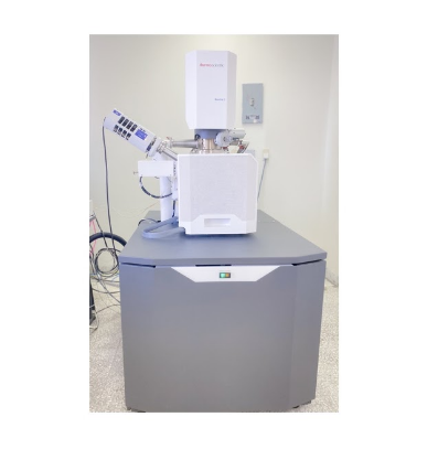
Field Emission Scanning Electron Microscopy
The Thermo Fisher Quattro S is an environmental scanning electron microscope(SEM) equipped with a Field Emission Gun. It is capable of operating at pressures up to50 mbar. The large pressure range allows the user to acquire images of hydratedspecimens. The low vacuum regime also enables users to take images of non-conductingsamples without coating their samples in gold or carbon.The Quattro S is equipped with an in-situ heating stage, which is capable of heating asample from room temperature up to 1400°C.
Main Features:
Resolution: 1 nm at 30 kV for SE, 2.5 nm at 30 kV for BSE.Acceleration voltage: 200 V to 30 kVPractical magnification: x6 to x250.000Detectors: ETD for SE, DBS for BSE signals, EDX detector for X-ray signals, LVD for low vacuum SE signals.Beam deceleration with stage bias: -4000 V to +50 VVacuum range: <1 x 10⁻⁴ Pa for high vacuum measurements and up to 100 Pa for low-vacuum measurements.Sample type: powders, films, metal plate (with size up to 2 cm wide and 0.5 cm thick)Suitable for analysis of nanomaterials, catalysis, metallic materials for corrosion studies, fracture and welding evaluation, projectile fragments, porous metals, metal foams, coating analysis, composite uniformity, graphite materials, and other carbon-based materials such as carbon papers, carbon fibers and activated carbons.Low vacuum mode (with water vapor) for humidity-dependent samples, polymer blends in composites, other non-conducting materials.

Location:
Chemistry department
Building #4, Room #157
Scientist In-Charge:
Name: Mr. Hassan Amasha
Phone: 3859
E-mail: hassan.amasha@kfupm.edu.sa
Scanning Electron Microscopy
Genesis-2120 is a high-performance W-filament SEM with x300K magnification and 3nmresolution. High performance and versatility are integrated in a stylish design which isthe most compact of the world in the same category. EmCrafts-Patented vacuum systemenables faster specimen exchange.
Main Features:
Resolution: 3 nm at 30 kV for SEAcceleration voltage: 200 V to 30 kVPractical magnification: x10 to x50.000Detectors: ETD for SE signals, and EDX for X-ray signals Vacuum: <9 x 10⁻³ PaSample type: powders, films, metal plate (with size up to 5 cm wide and 2 cm thick).Suitable for analysis of metallic materials for corrosion studies, fracture and welding evaluation, projectile fragments, porous metals, metal foams, coating analysis, composite uniformity, graphite materials, and other carbon-based materials such as carbon papers, carbon fibers and activated carbons.For non-conducting samples such ceramics, plastics, oxides, bacterial colony analysis and pharmaceutical materials appropriate gold or Pt coating is compulsory for SE imaging.