X-Ray Diffraction Laboratory
"We are committed to designing and developing original solutions for industry and society"
The Department of Chemistry's X-Ray Diffraction Facility Lab (XDFL) is equipped with one modern Bruker D8 Quest diffractometer, Bruker S8 TIGER WDXRF spectrometer, and Rigaku Ultima IV diffractometer. The single-crystal X-ray diffractometer is coupled to an extraordinarily sensitive Bruker AXS PHOTON II CPAD detector that gives rise to high-resolution data even on tiny crystals. Currently, the data collection can be performed at 298 K using ω-and φ-scans. The X-ray diffraction facility offers full data collection using APEX3 Crystal Structure Analysis Package (Bruker AXS). The X-ray Diffraction Facility provides a full single-crystal X-ray structure determination at MoKa (λ = 0.71073 Å) radiation generated by an Iμsmicrofocus tube.
Dr. Abdul Malik Peedikakkal
Supervisor
Mr. Nadir Osman
Supporting Staff
KFUPM
Bldg. 4 - Room 146
Dhahran, P.O. Box 5048
+966 13 860 2274
cls@kfupm.edu.sa
Sunday - Thursday
8:00 A.M - 4:00 P.M
KFUPM Faculty, Students, and Staff can utilize the facilities via booking through KFUPM RRM.
For outside KFUPM Community, you may book your reservations by registering at this link:
https://research.kfupm.edu.sa/RRM/Login.aspx
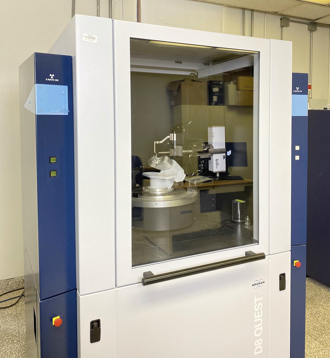
Bruker AXS D8 Venture diffractometer:
The X-ray Diffraction Facility offers a full single-crystal X-ray structure determination at MoKa (λ = 0.71073 Å) radiation generated by an Iμsmicrofocus tube. The single-crystal X-ray diffractometer is coupled to an extraordinarily sensitive Bruker AXS PHOTON II CPAD detector that gives rise to high-resolution data even on tiny crystals. The X-ray diffraction facility offers full data collection using APEX3 Crystal Structure Analysis Package (Bruker AXS).
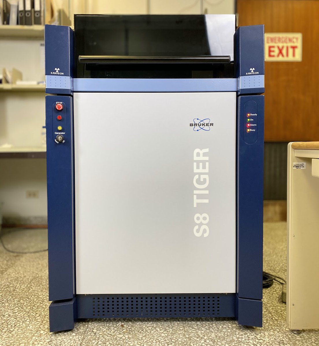
Bruker S8 TIGER WDXRF spectrometer:
Bruker S8 TIGER dispersive X-ray fluorescence (WDXRF) analysis provides the characteristics wavelength with a high degree of resolution. S8 TIGER with 3K provide high analytical performance with most flexible beam path.
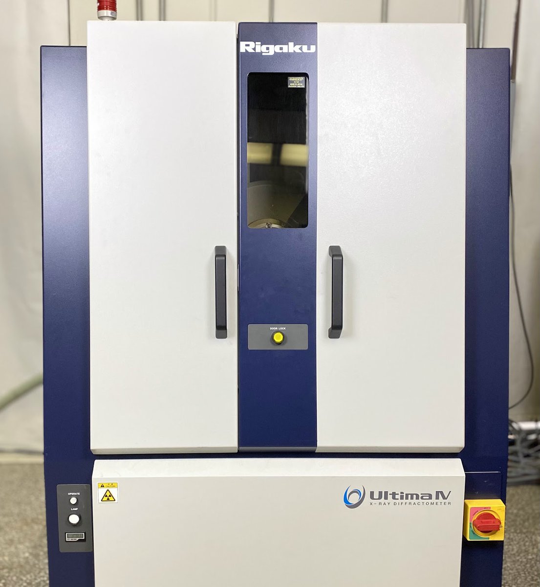
Rigaku Ultima IV diffractometer:
The Rigaku Ultima IV enables various applications, including in-plane and regular geometry phase identification, quantitative analysis, lattice parameter refinement, crystallite size, structure refinement, density, roughness, and multilayer thicknesses (from reflectivity geometries), and depth-controlled phase identification. Within the facility, users have access to the ICDD's PDXL2.
Bruker AXS D8 Venture diffractometer:
The X-ray Diffraction Facility offers a full single-crystal X-ray structure determination at MoKa (λ = 0.71073 Å) radiation generated by an Iμsmicrofocus tube. The single-crystal X-ray diffractometer is coupled to an extraordinarily sensitive Bruker AXS PHOTON II CPAD detector that gives rise to high-resolution data even on tiny crystals. The X-ray diffraction facility offers full data collection using APEX3 Crystal Structure Analysis Package (Bruker AXS).
Bruker S8 TIGER WDXRF spectrometer:
Bruker S8 TIGER dispersive X-ray fluorescence (WDXRF) analysis provides the characteristics wavelength with a high degree of resolution. S8 TIGER with 3K provide high analytical performance with most flexible beam path.
Rigaku Ultima IV diffractometer:
The Rigaku Ultima IV enables various applications, including in-plane and regular geometry phase identification, quantitative analysis, lattice parameter refinement, crystallite size, structure refinement, density, roughness, and multilayer thicknesses (from reflectivity geometries), and depth-controlled phase identification. Within the facility, users have access to the ICDD's PDXL2.
The Department of Chemistry's X-Ray Diffraction Facility offers:
Single-crystal X-ray diffraction
Only well-trained users will collect diffraction data at using Bruker D8 Quest diffractometer after approval. The crystal will be mounted on a MiTeGen™ loop in Paratone oil usually. Currently, the data collection can only be performed at room temperature. These diffractometers are coupled to extraordinarily sensitive Bruker APEX3 PHOTON II CPAD detector that gives rise to high-resolution data even on very small crystals.
Data collection will only be performed using Mo source, which provides higher resolution data.
Data collection using APEX3 and structural solution and cell refinement: SAINT, data reduction: SAINT, absorption correction: SADABS, structure solution: direct methods (SHELXT-2014) and structure refinement: full-matrix least-squares method on F2 (SHELXL-2014)5 using ShelXle as the graphical user interface, molecular graphics: program XP (part of the SHELXTL 6.14 program library) can be performed. The quality of data depends on the sufficient quality of crystal submitted.
Powder X-ray diffraction
Data can be collected for users, or trained users may collect their own data after the approval from the RRM. The Rigaku Ultima IV enables various applications, including in-plane and routine geometry phase identification, quantitative analysis, lattice parameter refinement, crystallite size, structure refinement, density, roughness, and multilayer thicknesses (from reflectivity geometries), and depth-controlled phase identification.
X-ray fluorescence spectroscopy
Whether it is qualitative or quantitative evaluation, SPECTRAplus leaves all options open: Scan measurements are always evaluated fully automatically, the elements are identified, and the concentrations are calculated. If you like, you can check and refine the results interactively. Data can be collected for users, or trained users may collect their own data after the approval from the RRM.
| Instrument | KFUPM First | KFUPM Additional | Non-Profit First | Non-Profit Additional | Commercial First | Commercial Additional |
|---|---|---|---|---|---|---|
| SXRD (Data Collection) | 350 | 250 | 225 | 195 | 600 | 520 |
| SCXRD (Structure Determination) | 300 | 250 | 450 | 375 | 1200 | 1000 |
| SCXRD (Unit Cell Determination) | 150 | 130 | 225 | 195 | 600 | 520 |
| XRF | 150 | 130 | 225 | 195 | 600 | 520 |
Description
Description
Location:
Chemistry Department
Phone: 94386
Building #4, Room #156
Scientist In-Charge:
Name: Mr. Nadir Osman
Phone: 4386
E-mail: nadirosman@kfupm.edu.sa
Powder X-Ray Diffraction
hen an X-ray is shined on a crystal, it diffracts in a pattern characteristic of the structure.In powder X-ray diffraction, the diffraction pattern is obtained from a powder of thematerial, rather than an individual crystal. Powder diffraction is often easier and moreconvenient than single crystal diffraction since it does not require individual crystals bemade. Powder X-ray diffraction (XRD) also obtains a diffraction pattern for the bulkmaterial of a crystalline solid, rather than of a single crystal, which doesn't necessarilyrepresent the overall material. A diffraction pattern plots intensity against the angle of thedetector, 2θ. An instrument dedicated to performing such powder measurements is called apowder diffractometer.Main Features:Fully automated optical alignment under computer control.Optional in-plane diffraction arm for in-plane measurements without reconfiguration.Focusing and parallel beam geometries without reconfiguration with CBO optics.D/teX Ultra 250 1D detector accelerates powder diffraction by a factor of 250 in speed and provides adjustable energy resolution ofapproximately 20% or 4% depending on sample type.SmartLab Studio-II is Rigaku’s full-function powder diffraction analysis package. Its modular design and automated flow bar user interface hasrevolutionized access to the power of XRD for the non-expert user.Various automated non-ambient stages are available. A low and medium elevated temperature (-180°C to 350°C) stage may be operated in air,gas, vacuum, or under liquid nitrogen cooling conditions.
Main Features:
Location:
Chemistry department,
Phone: 94386
Building #4, Room #156
Scientist In-Charge:
Name: Mr. Nadir Osman
Phone: 4386
E-mail: nadirosman@kfupm.edu.sa
Single Crystal X-Ray Diffraction
Single-crystal X-ray Diffraction is a non-destructive analytical technique which provides detailedinformation about the internal lattice of crystalline substances, including unit cell dimensions,bond-lengths, bond-angles, and details of site-ordering. Directly related is single-crystal refinement, wherethe data generated from the X-ray analysis is interpreted and refined to obtain the crystal structure.
X-ray diffractometers consist of three basic elements, an X-ray tube, a sample holder, and an X-raydetector. X-rays are generated in a cathode ray tube by heating a filament to produce electrons, acceleratingthe electrons toward a target by applying a voltage, and impact of the electrons with the target material.When electrons have sufficient energy to dislodge inner shell electrons of the target material, characteristicX-ray spectra are produced. These spectra consist of several components, the most common being Kα andKβ. Kα consists, in part, of Kα1 and Kα2. Kα1 has a slightly shorter wavelength and twice the intensity asKα2. The specific wavelengths are characteristic of the target material.
Main Features:
PHOTON II 7 CPAD technology
More than 2.5 times higher sensitivity for Mo radiation compared to HPC detectors.
Shutter less mode for unprecedented acquisition speed and data quality.
High intensity X-ray source for Mo- or Cu-radiation – no external cooling required.
Large enclosure with ample workspace.
APEX3 software suite – the must have for crystallography3-year warranty on PHOTON II 7 detector and X-ray tube.
Location:
Chemistry department,
Phone: 94386
Building #4, Room #156
Scientist In-Charge:
Name: Mr. Nadir Osman
Phone: 4386
E-mail: nadirosman@kfupm.edu.sa
X-Ray Fluorescence Diffraction
Instrument description
X-ray fluorescence (XRF) can directly analyze each element without destroying the sample. With XRF,measuring any type of solid or liquid is as easy as 1-2-3. In XRF, the sample is excited with a primaryX-ray beam, causing the sample to fluorescence. The primary X-rays eject electrons out of the inner atomicshells (K- and L-shell). The resulting “vacancy” is filled by an electron from an outer atomic shell. Thiselectron transition takes place only between the inner shells of the atom, which are not involved inchemical bonding . Due to the independency of chemical bonding, the samples can be analyzed directlywithout advanced sample preparation. This makes XRF the best method for elemental analysis.
How does XRF analyze elements? During electron transition, an electron drops from a higher to a lowerenergy atomic shell to fill the vacancy. The difference in energy is released as X-ray fluorescence radiation.This radiation has a characteristic wavelength for each element . XRF uses these different characteristicwavelengths or energies for elemental analysis.
Main Features:
The optimum level with respect to resolution, detection limits, and reliability are provided for each element.
The instrument has a high intensity X-ray tube and is combined with a compact beam path. It exhibits excellent excitation and high intensity over the wholeelemental range.
The selection of the analyzer crystal LiF 220 and the 0.23° collimator produces a choice combination for the analysis of traces from scandium to uranium.
Sample Care TM of the S8 TIGER offers distinct benefits to users. Since dust particles are a problem for instruments analyzing prepared samples as pressedpellets, sensitive spectrometer components are protected against being affected by sample particles with the 4x protection using a contamination shield.
The dust resistant design and sealed spectrometer cabinet dedicates the S8 TIGER for use in heavy industries such as ore processing and mining.
Location:
Chemistry department
Building #4, Room #157
Scientist In-Charge:
Name:
Phone:
E-mail:
Advanced Stereo Microscope
The Olympus SZX16 is designed for advanced research and leaves other stereos in its wake with amaximum numerical aperture (NA) of 0.3, producing a superior resolution of 900 line pairs per millimetre.With such amazing resolution (and peerless magnification) you can make your work more efficient, moreprecise and gain much more information from your samples. The Olympus SZX16 provides a large zoomratio of 16.4:1. With this seamless zoom ratio combined with the most comprehensive range of parfocalobjectives (0.5x, 1.0x, 1.6x & 2.0x), the SZX16 can take you from a macro-view to a micro-view allowinge.g. visualisation of ultra precise details on manufactured micro devices
Main Features:
- A wide zoom range between 0.7x and 11.5x allows the user to observe fine details while maintaining a wide field of
view without replacing objective lenses.
- Large Selection of Objective Lenses
- The SZX16 motorized focus drive makes digital documentation with extended focal imaging (EFI) efficient and fully automatic.